

10 DVT (deep vein thrombosis) facts
In this article we will describe 10 in depth facts about 10 DVT (deep vein thrombosis). This article is mainly for health professionals.
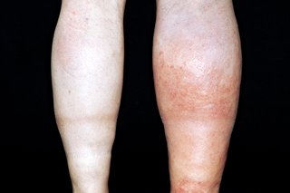
Left DVT. This patient also has varicose eczema.
Key Points
- Definition: DVT is a blood clot in a deep vein of an extremity (usually calf or thigh) or the pelvis. DVT is the primary cause of pulmonary embolism
- Causes: conditions that impair venous return, leading to endothelial injury or dysfunction, or hypercoagulability
- Symptoms: DVT may be asymptomatic, or cause pain and swelling usually in a calf or thigh
- Diagnosis: is by history and examination and is confirmed by testing with duplex ultrasonography. A negative D-dimer helps to exclude DVT, whereas a positive result is nonspecific and requires additional testing to confirm DVT
- Treatment: is with anticoagulants.
1. Definition
- Deep venous thrombosis (DVT) refers to the formation of a blood clot in a deep vein, most commonly in the calf, thigh, or pelvis
- Venous thromboembolism (VTE) is a broader term that includes both DVT and pulmonary embolism (PE).
2. Epidemiology
- Untreated DVT: 20% of individuals with DVT will develop a pulmonary embolism.
- Concurrent PE: 45-50% of patients with DVT may have a concurrent PE.
- Cardiovascular Disease: In the UK, DVT and PE together form the third most common cardiovascular condition, following myocardial infarction and stroke.
3. Risk factors
- Age (especially >60 years)
- Obesity and smoking
- Cancer and heart failure
- Varicose veins or genetic predispositions
- Pregnancy, oral contraceptives (OC), hormone replacement therapy (HRT)
- History (or family history) of DVT or PE
- Prolonged immobility (e.g. long flights, bed rest).
4. Causes
- Clotting disorders (e.g. Factor V Leiden, Antiphospholid antibody syndrome)
- Injury to a vein (e.g. trauma, surgery)
- Certain medications
- Extended periods of immobility (e.g. hospitalisation, long travel)
5. Symptoms
- Swelling, pain, tenderness, and redness in the affected area (usually the calf, sometimes the thigh)
- Some individuals may be asymptomatic
- DVT can also occur in the arm or abdomen, leading to similar symptoms in those regions.
6. Diagnosis
DVT diagnosis is usually confirmed through:
- Imaging tests: Ultrasound is most common, but venography can be used if necessary.
- Blood tests: D-dimer test, which has high sensitivity but low specificity.
DVT shown on venogram
D-dimer
- A high D-dimer may indicate clot formation but can be raised in other conditions (e.g. pregnancy, malignancy, AKI, CKD, infection, recent trauma, or surgery).
- Key point: A negative D-dimer has high negative predictive value and can effectively rule out DVT (and PE).
Differential diagnosis
- Soft-tissue trauma
- Cellulitis
- Compression of pelvic veins or lymphatics
- Ruptured popliteal cyst (Baker’s cyst)
- Unilateral oedema.
7. Treatment
The primary treatment for DVT is anticoagulation to prevent clot extension and new clot formation. Options include:
- Heparin – e.g. low-molecular-weight heparin
- Warfarin or
- Direct-acting oral anticoagulants (DOACs) – e.g. apixaban, rivaroxaban.
In severe cases, clot removal or thrombolytic therapy may be required.
Pregnancy: Pregnant patients are typically treated with subcutaneous heparin throughout pregnancy and for 6 weeks postpartum.
8. Complications
- The most serious complication of DVT is pulmonary embolism (PE), where the clot dislodges and travels to the lungs, potentially causing fatal outcomes. Without treatment, 10-30% of DVT patients will develop PE
- Post-thrombotic syndrome (PTS) is a long-term complication of DVT, involving chronic pain, swelling, and skin changes in the affected limb due to venous damage.
9. Prognosis
- With timely and appropriate treatment, the prognosis for DVT is generally good
- Most clots dissolve naturally over time with anticoagulant therapy preventing new clots from forming.
10. Prevention
- Staying active and avoiding prolonged immobility
- For high-risk patients, wearing compression stockings
- For high-risk patients or those with recurrent DVT, anticoagulation therapy (e.g. warfarin, DOACs) may be necessary
- In certain cases, an inferior vena cava (IVC) filter may be inserted to prevent PE in patients with recurrent DVTs.
Lifestyle adjustments: Patients are advised to stop smoking, maintain a healthy weight, and increase physical activity to reduce the risk of recurrence.
Summary
We have described 10 in depth facts about DVT (deep vein thrombosis). We hope it has been helpful.
Other resources
PE facts: in depth
DVT (NHS England)
DVT (NHS Scotland
Review: Waheed, 2023
