A normal chest x-ray – the basics
In this article we will describe what is a normal chest x-ray and how to interpret it.

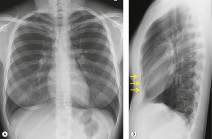
This is a normal chest x-ray (CXR). It is a PA and lateral chest x-ray of a healthy woman. Note mild pectus excavatum on the lateral image (arrows).
Why is this a normal chest x-ray?
This is a normal chest x-ray because it has the following features:
- Normal bones and soft tissues
- Clear lung fields
- Normal looking heart (CT ratio under 0.5)
- No visible nodules, tumours, masses or foreign bodies
- Clearly outlined chest cavity, with clear costodiaphragmatic angles
- Normal vasculature.
Let’s go into some more detail.
Features of a normal chest x-ray
A normal chest X-ray typically displays the following features:
Bony Structures
-
Clavicles: Symmetrical and intact
-
Scapulae: Visible, with normal contours
-
Ribs: 10-12 pairs, with normal curvature and no fractures
-
Thoracic spine: Visible, with normal alignment and no fractures.
Soft Tissues
-
Lung parenchyma: Uniformly translucent, with no nodules or consolidations
-
Pleura: Not visible or minimally visible, indicating no pleural effusion
-
Mediastinum: Central, with normal width (less than 8 cm)
-
Heart: Normal size and shape, with a cardiothoracic ratio less than 50%
-
Diaphragm: Dome-shaped, with normal position and movement.
Pulmonary Vasculature
-
Pulmonary arteries: Visible, with normal calibre and no dilation
-
Pulmonary veins: Visible, with normal calibre and no dilation.
Other Features
-
Trachea: Central, with normal calibre and no deviation
-
Bronchi: Visible, with normal calibre and no dilation
-
Costophrenic angles: Sharp, indicating no pleural effusions.
Why are chest x-rays done?
A CXR is quick, cheap and non-invasive – and tells you alot about the heart (like an ECG) and lungs. This is why such an ‘old test’ is still so widely used.
Summary
We have described the normal chest x-ray. We hope it has been helpful.

