

Basic principles of x-ray analysis
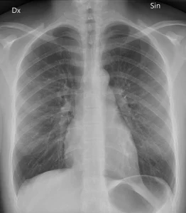
A normal chest x-ray
The interpretation of an x-ray film requires sound anatomical knowledge, and an understanding that different tissue types absorb x-rays to varying degrees:
- High density tissue (e.g. bone) – absorb x-rays to a greater degree, and appear white on the film.
- Low density tissue (e.g. lungs) – absorb x-rays to a lesser degree, and appear black on the film.
- Intermediate density tissue (e.g. muscle and fat) – appears as shades of grey on the x-ray film.
It is important to appreciate that x-rays only give a 2D superimposed view of the body part that has been imaged.
Therefore, it may be necessary to take views of the same area from different angles, e.g.
- In cases of suspected fracture, to gain a full understanding of the injury.
- In pneumonia, to decide exactly where is the consolidation (a lateral film is then done).
Comparison to other imaging techniques
The biggest advantage with plain film x-rays is the amount of radiation involved. It offers lower dosage compared to CT, and certain studies are performed relatively quickly (chest x-rays).
They are often used as an initial screening to rule out anything obvious before an advanced modality is used such as CT or MRI.
However, plain film x-rays procedures are being replaced by CT and MRI due to advancements in technology.
There are CT scanners available on the market now that offer radiation dosage levels as low as plain film x-rays.
Below is a summary table of the common imaging modalities.
| Factor | CT | MRI | X-ray | Ultrasound |
| Duration | 3-7 minutes | 30-45 min | 2-3 min | 5-10 minutes |
| Cost | Cheaper | Expensive | Cheap | Cheap |
| Dimensions | 3 | 3 | 2 | 2 |
| Soft tissue | Poor detail | Excellent detail | Poor detail | Poor detail |
| Bone | Excellent detail | Poor detail | Excellent detail | Poor detail |
| Radiation | 10mSv | None | 0.15mSv | None |
Depending on the tissue being imaged, the urgency of the investigation and the level of detail required, any of these techniques may be preferred.
