

Chest x-ray in pneumonia
On a chest x-ray, pneumonia appears as an area of increased opacity or whiteness, indicating lung consolidation where air is replaced by fluid or pus. Here are some examples.
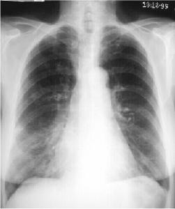
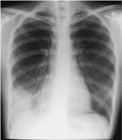
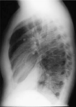
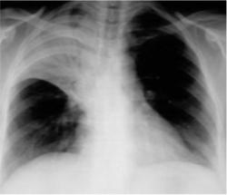
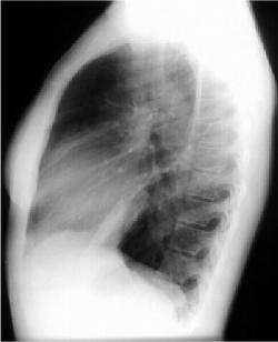
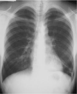
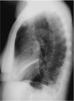 Right Middle Lobe Consolidation
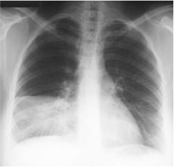
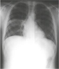
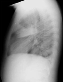
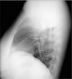
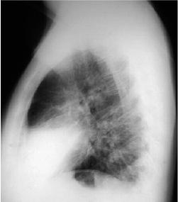 Right Middle Lobe Pneumonia
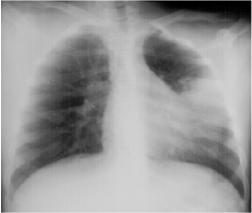
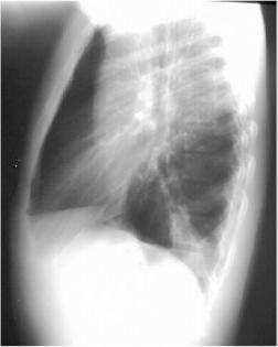
 Right Lower Lobe Pneumonia
Right Lower Lobe Pneumonia, Anterior Segment
 Right Lower Lobe Pneumonia, Superior Segment
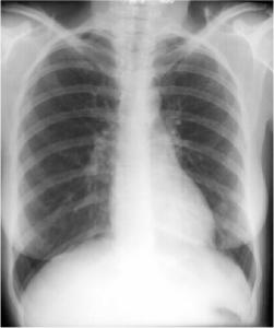
 Right Upper Lobe Pneumonia
 Left Lingular Pneumonia
 Left Lower Lobe Pneumonia, Anterior Segment
 Left Lower Lobe Pneumonia, Posterior Segment
