

What is a chest xray – and how is it done?
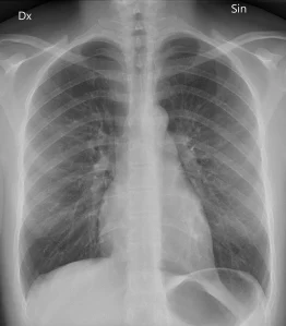 A normal chest x-ray (CXR)
In this article we will describe what is a chest xray – and how it is done.
A chest x-ray is a painless non-invasive medical imaging test used to take pictures of the lungs, heart, and surrounding tissues. Here’s a step-by-step guide on how it is done:
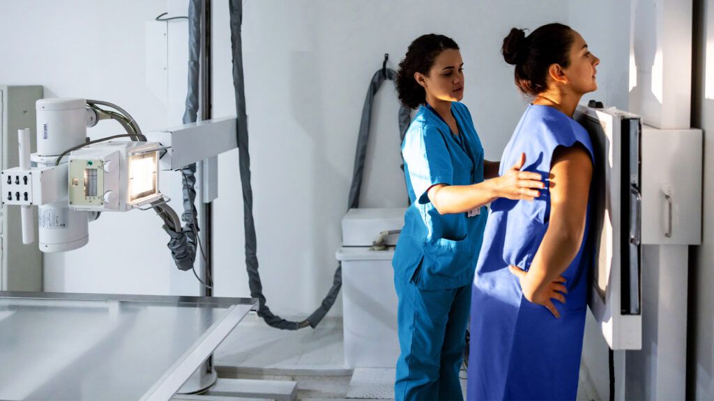
Preparation
-
You will be asked to remove any clothing or jewellery from your upper body
-
You may be given a hospital gown to wear
-
Inform the technician (called a radiographer) if you are pregnant or have any medical implants.
Positioning
-
You stand (usually) or sit in front of the x-ray machine
-
Your chest will be positioned against the image receptor (a digital plate or film).
-
Your arms will be raised above your head or to your sides.
Taking the x-ray
-
Hold your breath and remain still for a few seconds
-
The technician will activate the x-ray machine, which will emit a low-level radiation beam
-
The beam passes through your chest, creating an image on the digital plate or film.
The x-ray plate is touching the patients chest, with the x-rays coming from behind (called ‘posterior’).

Views
-
Typically, two views are taken (as shown above):
-
Posteroanterior (PA) view: taken from the back to the front
-
Lateral view: taken from the side.
-
After the test
-
The technician will review the images for quality
-
You will be free to go once the test is complete
-
A radiologist (x-ray doctor) will interpret the images and send the results to your doctor.
Remember, a chest x-ray is a quick and painless test that helps doctors diagnose and monitor various medical illnesses, such as lung diseases (including cancer), and heart conditions.
Summary
In this article we have described what is a chest xray, and how it is done. We hope it has been helpful.
