How to make diagnoses from nail abnormalities
All of the conditions below can be diagnosed through the nails, with a bit of experience.
Nail abnormalities include variations in the shape, colour, thickness, and texture of the fingernails, toenails, or both. Some changes are normal and harmless; whilst others may be a sign of injury, infection, or an acute/chronic medical condition that requires medical attention.
Acral lentiginous melanoma (involving nail)
Whilst acral lentinginous melenoma is often seen anywhere on the palms, soles, and even in the mouth. When it occurs within the nail, a clue that this is melanoma is involvement of the periungal regions as seen in this picture.
Beau’s lines
Transverse depressed ridges seen in severe infection, MI, hypotension/shock, hypocalcemia, post-surgical, malnutrition and with certain chemotherapy. It can follow COVID-19 as well.
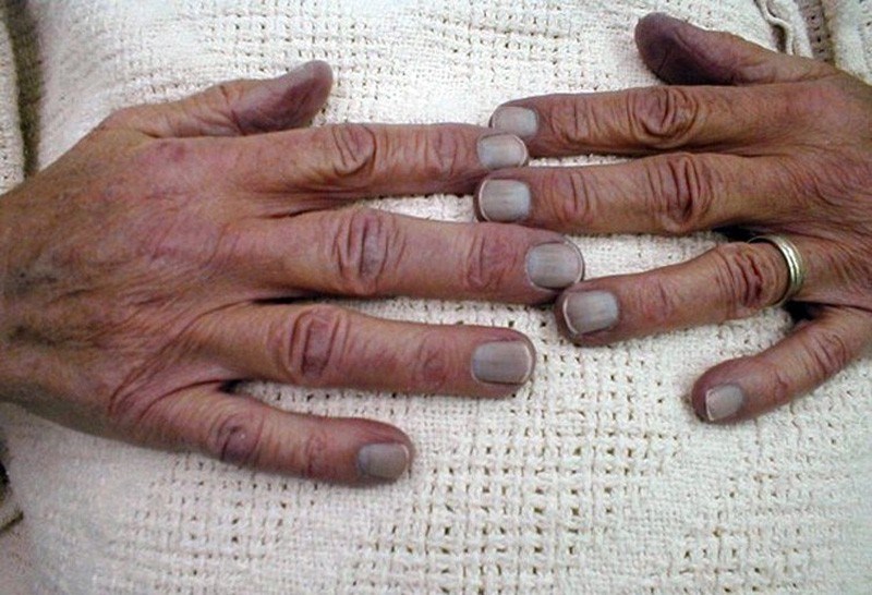
Blue half-moon nails

This can be due to poisoning. The picture above is in someone with silver poisoning (called agyria or argyrosis; and of face).
Blue nails (cyanosis)
Blue nails can be a symptom of cyanosis, which is a condition where the skin or mucous membranes appear bluish due to a lack of oxygen in the blood.
Brittle nails

Brittle nails are common. Here are some causes.
- Ageing: Brittle nails are a common result of aging.
- Chronic medical conditions: Iron or zinc deficiency, thyroid disease, anaemia, or anorexia nervosa/bulimia
- Nail habits: Overuse of nail polish, manicures, nail polish remover, harsh soaps, and detergents
- Trauma: Repeated trauma to the nails can cause splitting and softening.
- Wetting and drying: Repeated wetting and drying of the hands can make nails expand and shrink, leading to brittleness. This is often worse in low humidity and in the winter.
Clubbing (also known as ‘drumstick fingers’ or Hippocratic nails)
This patient has a nail-fold infarct as well, suggesting possible infective endocarditis.

Clubbing of the nails is soft tissue swelling of the terminal phalanx, resulting in the straightening of the angle between the nail bed and the nail. The angle between nail plate and proximal nail fold is greater than 180 degrees.
Causes of clubbing include:
- Respiratory
- Lung cancer. Clubbing is in general an ominous sign for this, and remember ‘beware of the yellow clubbed digit’ (yellow from nicotine, and clubbed from cancer)
- Suppurative lung disease (bronchiectasis, cystic fibrosis, lung abscess and empyema)
- Pulmonary fibrosis (and its many causes)
- Asbestosis/pleural mesothelioma
- COPD IS NOT A CAUSE OF CLUBBING (if you seen clubbing in a COPD patient, think lung cancer).
- Cardiac
- Congenital cyanotic heart disease (L to R shunts)
- Infective endocarditis
- Atrial myxoma.
- Gastrointestinal
- Inflammatory bowel disease
- Primary biliary cirrhosis
- Coeliac disease (and other cause of chronic malabsorption)
- GI lymphoma.
- Other
- Thyrotoxicosis (thyroid acropachy)
- Hereditary.
There is a longer list of the 50+ causes of clubbing here on MyHSN.
Defluvium unguium (onychomadesis; complete loss of nails)

Certain systemic diseases such as scarlet fever, syphilis, leprosy, alopecia areata,ulcerative colitis (patient above) and exfoliative dermatitis; dermatoses such as nail infection, toenail psoriasis, eczema; arsenic poisoning.
Ingrown toenail
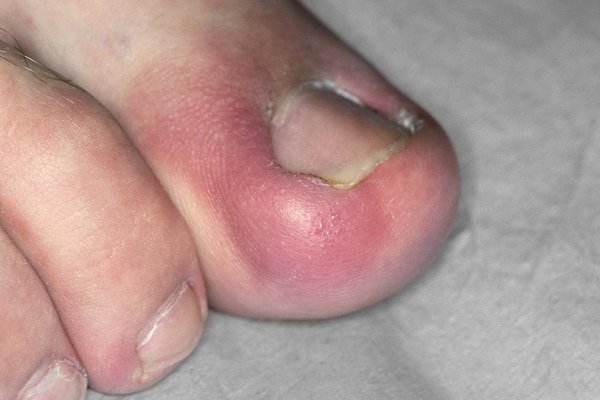
An ingrown toenail is a common problem where the nail grows into the toe.
Koilonychia (‘spoon’ nails)
:max_bytes(150000):strip_icc():format(webp)/VWH-DermnetNZ-Koilonychia-Spooning-01-669aac78a66644eb8b6d1d125541766b.jpg)

This is a deformity of the nails where the central portion of the nail is depressed and the lateral aspects of the nail are elevated. It can be due to chronic iron deficiency anaemia, secondary to malnutrition, worms, coeliac disease, gastrointestinal blood loss, and malignancy.
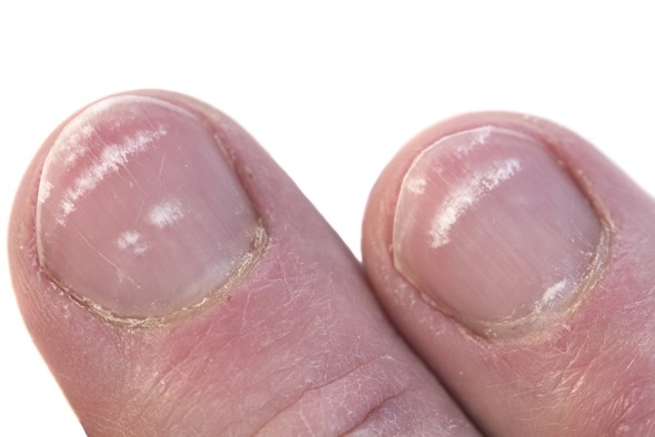
Leukonychia
This is most commonly caused by minor injuries, such as nail biting, or may occur while the nail is growing. It also may be caused by calcium deficiency, and hypoalbuminaemia of chronic liver disease (see below).
Lindsay’s nails (‘half-and-half nails’)

Distal brown transverse band seen in chronic kidney disease (CKD). Caused by increased pigment deposition.
Mees’ lines

Transverse while lines (usually one per nail, no depressions) that often can will disappear if pressure is placed over the line. It is associated with arsenic, thallium poisoning and other heavy metal poisoning.
Muehrcke’s lines (leukonychia striata)
:max_bytes(150000):strip_icc():format(webp)/VWH-DermnetNZ-Leukonychia-MeesLines-01-1bee98819c024f3c9e5cd478a5799730.jpg)
Narrow while transverse lines (not depressed, compared to Beau’s lines). Usually 2 or more lines on one nail. Seen in states of decreased protein synthesis or increased protein loss, such as with hypo-albuminaemia (usually less than < 20 gL), certain chemotherapy and nephrotic syndrome.
Nail pitting
Non-specific sign for psoriasis (additional signs include onycholysis, thickening, and ‘oilspot’ lesions which are yellow patches on the nail).
Nailfold infarcts
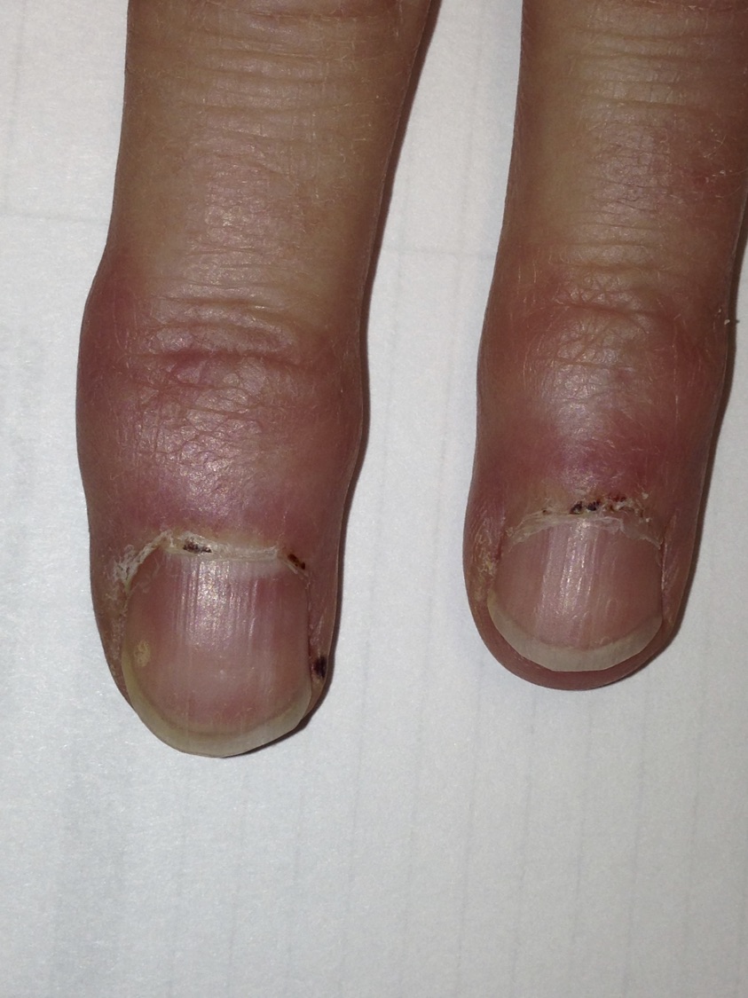
Seen in infective endocarditis and many autoimmune diseases.
Nicotine stained nails

Nicotine stained distally, but not proximally with clear line of demarcation. May also appear when patient switches to ‘lower tar’ tobacco.
Onychogryphosis (ram’s horn nails)
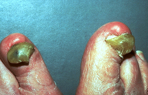
This happens when the nails thicken and overgrow. It is usually age-related but can be genetic as well.
Onycholysis (nail lifting up)
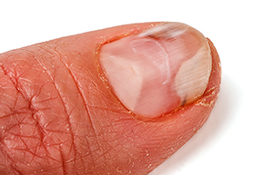
There are several causes:
- Fungal infection
- Ageing
- Psoriasis
- Injury from an aggressive manicure
- Injury form cleaning under nails with a sharp object.
Onychotillomania (washboard nails)
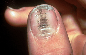
This means grooves and ridges in the centre of the thumb, due to a habit of picking at (or pushing back) the cuticles on thumbnails. Many people are unaware that they do this.
Onychomycosis

A fungal nail infection. There are various types. Above is superficial white onychomycosis, typically caused by T. interdigitale or
C. albicans.
Paronychia

Inflammation of the nail folds – red, swollen, often tender. Frequent immersion in water a risk factor for chronic paronychia.
Red-violet half moon bands

Clinical manifestations of the patient with systemic lupus erythematosus (SLE, lupus). The red-violet half-moon-shaped bands were present in all fingernails except left small finger. Note violaceous rashes on ears.
Splinter haemorrhages

Nonspecific finding associated with trauma most commonly but also seen in infective endocarditis and many autoimmune diseases.
Subungual haematoma
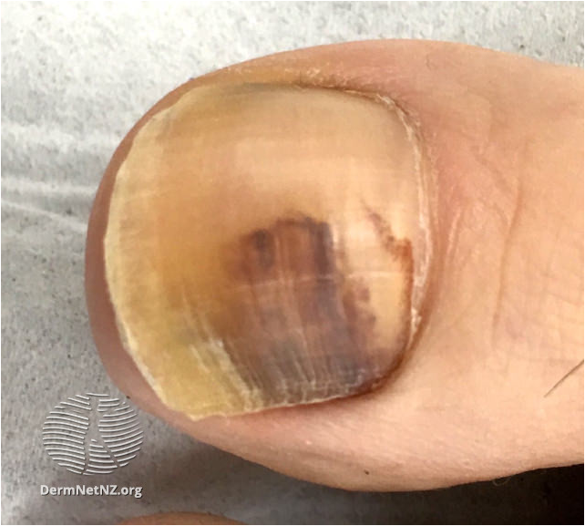
History of trauma (though may not be remembered). Discolouration migrates with nail growth, with clearance of proximal nail bed with time. Discolouration is usually red or red/black but not brown. No treatment is required.
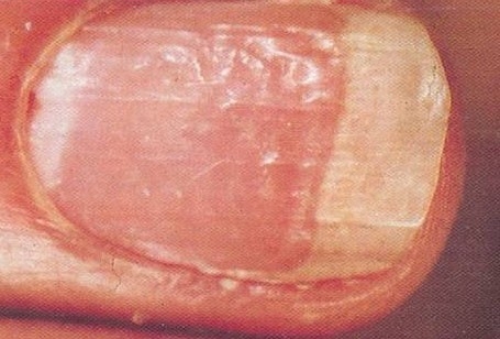
Terry’s nails
:max_bytes(150000):strip_icc():format(webp)/VWH-DermnetNZ-TerryNails-01-35ca03034b6e42c99dca55301a1f2a1e.jpg)
Proximal paleness extending halfway up the nail, often eliminating the lunula. Darker distal band. Seen in states of stress (e.g. advanced age, chronic liver disease/cirrhosis, CHF, DM2).
Yellow nails
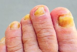
There are many causes. Most are due to fungal infections, ageing or both.
- Fungal infections
- Onychomycosis: a fungal infection that penetrates the nail and causes discoloration. Usually the first (big) toenail is affected first.
- Ringworm: a fungal infection that can spread to the nails.
- Ageing. As people age, their nails become thicker, more brittle, and more prone to discolouration.
- Other causes
- Psoriasis: usually associated with nail pitting.
- Nail polish: using nail polish that contains harsh chemicals or dyes can stain nails yellow.
- Genetics: some people may be more prone to yellow nails due to their genetic makeup
- Yellow nail syndrome: A rare disorder that can cause yellow nails, along with respiratory symptoms and lower limb lymphoedema. The cause is unknown, but it may be genetic or acquired.
- Chronic medical conditions
- Diabetes: high blood sugar levels can cause nail discoloration.
- Chronic liver disease: liver dysfunction can lead jaundice (that includes the nails).
- Chronic kidney disease (CKD) and/or nephrotic syndrome: kidney dysfunction can cause a buildup of waste products, leading to nail discoloration.
- Bronchiectasis.
- Thyroid disease: hypothyroidism (underactive thyroid) or hyperthyroidism (overactive thyroid) can cause nail changes.
- Rheumatoid arthritis (RA).
- Lifestyle factors
- Smoking: nicotine and tar in cigarettes can stain nails yellow.
- Excessive exposure to chemicals: detergents, cleaning products, or nail polish removers can cause nail discolouration.
- Poor diet: lack of essential nutrients, vitamins, and minerals can affect nail health.
Summary
We have described many nail abnormalities; and the conditions that can you diagnose when you see them. We hope it has been helpful.
Other resources
Review article: Lee, 2022
Nail diseases


