What happens to a biopsy after it has been taken?
Once your biopsy has been taken it will go to the pathology laboratory. The biopsy will be processed in the laboratory to stain the tissue and to cut it into very thin slices.
These are then looked at under a microscope by a pathologist. A pathologist is a doctor that specialises in making a diagnosis of disease in tissue samples. This means they use a microscope to look at your biopsy in detail, to work out exactly what it is going on.
The pathologist writes a report that your doctor or nurse can access and then discuss with you. The pathologist may also discuss your biopsy case at a Multidisciplinary Team meeting (MDT), so the whole team can decide on the best treatment.
It can sometimes take time (up to several weeks) to work out what your biopsy shows, and extra tests may be needed. In fact, sometimes they are sent to other hospitals for more specialist analysis. But if you are worried or there is a delay, speak to your doctor or nurse.
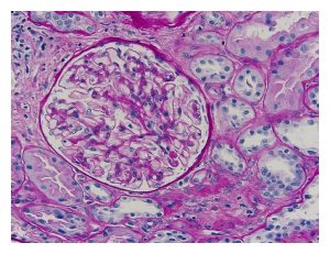
What does a biopsy look like down the microscope?

This is a normal kidney biopsy. The circular thing towards the left is a glomerulus, the filtering unit of the kidney. There are a million in each kidney (you have two). Many biopsies look a bit like this, and are pink after staining.

