What is a normal chest x-ray?
In this article we will describe what is a normal chest x-ray and how to interpret it.

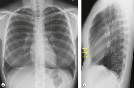
This is a normal chest x-ray (CXR). It is a PA and lateral chest x-ray of a healthy woman. Note mild pectus excavatum on the lateral image (arrows).
A CXR is quick, cheap and non-invasive – and tells you alot (like an ECG). This is why such an ‘old test’ is still so widely used.
How to read a CXR
You need to know your anatomy.

Then there are 3 stages.
1. Determining the view
- Posteroanterior (PA; normal): patient stands or sits upright approximately 6 feet in front of the beam, with the x-ray film on the front of the chest, and hands behind the head

- Lateral: patient stands or sits upright with his or her arms raised and turns on to left side, facing the film
- Anteroposterior (AP): patient sits or lies down on top of the film, and the x-ray beam travels through the patient from front to back.
2. Determining the quality
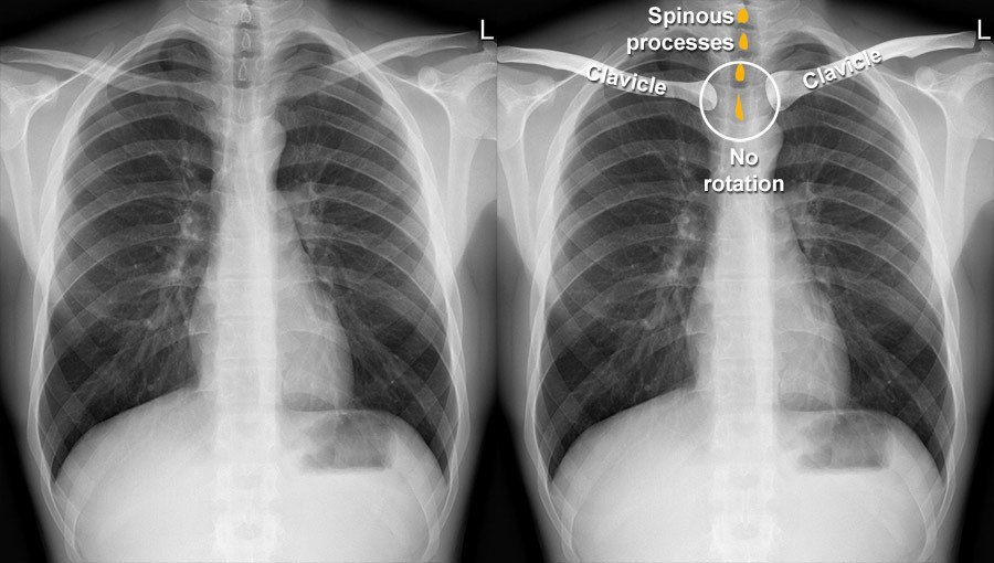
- Rotation: compare the positions of the left and right medial clavicular joints to the spinous processes. There should be an equal gap on each side, as shown in the following diagram

- Inspiration: count the ribs visible in the lung fields. There should be 10 posterior (8 is minimum) and 6 anterior ribs

- Penetration: the vertebrae behind the heart are just visible.
MNEMONIC – RIP
3. Systematic analysis
- Air (lung fields; compare R and L in zones; opacities?); airway (trachea, central?); apices (TB?)
- Bones (clavicles; ribs (fractures?); breasts (male vs female; are there 2 breasts?)
- Cardiac shadow; carina (visible)
- Cardiothoracic ratio (CTR) is measured on a PA chest x-ray, and is the ratio of maximal horizontal cardiac diameter to maximal horizontal thoracic diameter (can only comment on CTR on a AP)
- A CTR greater than 0.5 is abnormal ( i.e. heart size should be <50% of thoracic width)
- Enlarged heart (cardiomegaly; heart failure, hypertrophy, or cardiomyopathy?)
- Diaphragm (anything in the costodiaphragmatic angle, i.e. effusion?)
- Extrathoracic soft tissues (outside the bones); effusions; edges (look down sides of lungs, pneumothorax?)
- Foreign bodies (central line, pacemaker, ET or NG tube?)
- Gastric bubble (and gas under R hemi-diaphragm, i.e. perforation?); great vessels
- Hilum
- Impression (conclusion, ready to present; see below).
MNEMONIC – Alphabet (ABCDEFGHI)
Note. ‘This A-I’ mnemonic is not the best order to do it in. This order is better ..
V – view (PA or AP?)
Q – quality of film
E – bones and soft tissues (including breasts)
F – foreign bodies
A – apices
A – air (lungs)
C – cardiac shadow
D – diaphragm and costodiaphragmatic angles (and under R hemi-diaphragm)
G – gastric bubble
H – hilum
I – impression.
How to present
“This is a PA chest x-ray of Mr/Mrs X/Y. The heart is of normal size with a CTR of under 0,5. The lungs are clear. It is normal”
Or
“This is an AP chest x-ray. I therefore cannot comment on the heart size. There is increased shadowing in the R lower zone. The most likely diagnosis is pneumonia, but the differential includes pulmonary haemorrhage and embolism”.
Summary
We have described what is a normal chest x-ray and how to interpret it. We hope it has been helpful.

