What are the different types of x-ray?
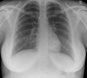
 This is a normal chest x-ray
In this article will explain what is an x-ray.
Key Points
- An x-ray is a painless and quick procedure commonly used to produce images of the inside of the body.
- An x-ray uses a small amount of radiation.
- X-rays are used to diagnose disease and injury.
- A contrast dye, such as iodine or gadolinium, is used during some x-rays to help to improve images.
- Before an x-ray you should tell the doctor if you are pregnant or may be pregnant.
It is a very effective way of looking at the bones and can be used to help detect a range of conditions.
X-rays are usually carried out in hospital x-ray departments by trained specialists called radiographers; although they can also be done by other healthcare professionals, such as dentists.
How x-rays work
X-rays are a type of radiation passes through the body. They cannot be seen by the naked eye and you can’t feel them.
As they pass through the body, the x-rays are absorbed at different rates by different parts of the body. A detector on the other side of the body picks up the x-rays after they have passed through and turns them into an image.
Dense parts of your body that x-rays find it more difficult to pass through, such as bone, show up as white areas on the image. Softer parts that x-rays can pass through more easily, such as your heart and lungs, show up as darker areas.
What are the types of x-rays?
There are several different types of x-ray:
- Plain radiography (plain x-ray)
- Computed tomography, known as a ‘CT scan’
- Fluoroscopy — which produces moving images of an organ
- Mammography — an x-ray of your breasts
- Angiography — an x-ray of your blood vessels
- Bone density scan — helps identify low bone density and diagnose osteoporosis.
What are x-rays used for?
X-rays are used to examine most areas of the body. They are especially used to look at bones and joints. Although they are also used to detect problems affecting soft tissue, such as internal organs.
Problems that may be detected during an x-ray include:
X-rays are used to diagnose disease and injuries, including:
- Bone conditions — such as fractures, dislocations, bone infections, arthritis and osteoporosis
- Lung conditions — such as pneumonia and collapsed lung
- Heart failure
- Kidney stones
- Blood vessel problems — such as an aortic aneurysm (a bulge in the aorta)
- Cancer — such as lung cancer, bone cancer and breast cancer
- Blockages in your bowel
- Tooth problems, including decay, loose teeth and dental abscesses
- Detection of foreign objects — such as when a child accidentally swallows an object
- Scoliosis (abnormal curvature of the spine)
- To check the position of wires, leads and tubes after a surgical procedure.
How an x-ray is done

This is how a chest x-ray is done. The patient stands in front of a plate that detects the x-rays passing through the body from behind.
Do x-rays have any risks?
Few. The occasional ‘simple’ chest or bone x-ray has almost no risks. There are small risks to some of the more complex x-rays (like an angiogram or CT scan). The addition of dye increases the risk. The dye can damage the kidneys and produce temporary (that reverses) or permanent damage. So such x-rays should be done with caution in people with CKD.
Summary
In this article we have described what are the different types of x-ray. We hope it has been helpful.

